 |
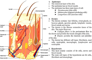
| Structure & Function of Skin |
The skin serves many purposes:
- serves as a barrier to the environment, and some glands (sebaceous) may have weak anti-infective properties.
- acts as a channel for communication to the outside world.
- protects us from water loss, friction wounds, and impact wounds.
- uses specialized pigment cells to protect us from ultraviolet rays of the sun.
- produces vitamin D in the epidermal layer, when it is exposed to the sun's rays.
- helps regulate body temperature through sweat glands.
- helps regulate metabolism.
- has esthetic and beauty qualities.
Skin Layers
The skin is composed of several layers. The lowest layer is called the dermis. This layer is composed of connective tissue, blood vessels, nerve endings, hair follicles, and sweat and oil glands.
The outermost or top layer of skin is called the epidermis. This is the layer of skin which we see. This layer rests on top of the dermis.
The thickness of the epidermis varies with your age, your sex, and the location on the body of the skin. For example, the epidermis on the underside of the forearm is about 5 cell-layers thick. On the sole of the foot, the epidermis might be 30 cell-layers thick. The epidermis is for the most part impermeable to water. This water-impermeable skin is one of the features that allows us to live on land.
Several cell types are present in the epidermis. The epidermis consists of many layers:
- The stratum corneum or outer layer. This layer is made of flattened epithelial cells in multiple layers. These layers are called keratinized layers because of the build-up of the protein keratin in those cells. Keratin is a strong protein that is specific to the skin, hair and nails. This layer of skin is, for the most part, dead--it is composed of cells that are almost pure protein.
- The translucent or transitional layer. This is a translucent, thin layer of cells. This layer is sometimes visible in thick skin; however, nuclei and other organelles are not visible. The cytoplasm (the amorphous area between the nucleus and the outer membrane of the cell) is mostly made of keratin filaments.
- The suprabasal layers. This is three to five layers of flattened polygonal cells that have granules in the cytoplasm. Below them is a layer of cube-shaped cells that also contain bundles of keratin filaments.
- The basal or cell-division layer. This layer is just above the basement membrane and the dermis. It is a single layer of cells that undergo cell division to renew the upper layers of the epidermis.
The human epidermis is renewed every 15-30 days. In some disease states it is renewed in about 7-10 days (e.g. psoriasis).
 |
| skin structure and function |
Skin Cell Types
Keratinocytes
The most abundant cell type of the epidermis is the keratinocyte. These cells produce keratin proteins that provide some of the rigidity of the outer layers of the skin. Keratinocytes also form the bulk of the material in hair follicles. Dandruff and hair are dead keratinocytes.
Fibroblasts
The dermis is produced largely by fibroblasts, which during embryonic development are part of the mesenchyme. The fibroblasts produce the collagens and elastins that make skin very durable, from within.
Melanocytes
Melanocytes are cells in low abundance in the epidermis that produce the pigment melanin. The pigment made in melanocytes is transferred to the cells of the hair or epidermis. The melanin granules are injected into (or ingested by) the keratinocyte cells. There, the melanin granules accumulate around the nucleus of each keratinocyte.
Melanin absorbs harmful ultraviolet (UV) light before the UV radiation can reach the nucleus. Melanin protects the DNA in the nucleus from UV radiation damage. When melanin is produced and distributed properly in the skin, dividing cells are protected from mutations that might otherwise be caused by harmful UV light.
Differences in skin color are due mostly to differences in the types and amount of pigment in our keratinocytes. Skin darkening (tanning) from sun exposure is caused by the movement of existing melanin into keratinocytes, and by increased production of melanin by the melanocyte.
During embryonic development these cells migrate from the neural crest into the skin.
Langerhans cells
These are star-shaped resident immune cells, macrophages. A macrophage is a cell that protects your body from injury or illness. Macrophages break up or destroy (phagocytise) the invading organisms. These macrophages process the invading organisms and present antigens to the T-lymphocytes. The T-lymphocytes are immune-system cells which ultimately identify a substance as foreign or dangerous to the body.
Merkel's Cells
Only a few of these cells are present in skin; they are more numerous in the palms and soles (feet). These cells are probably sensory mechanical receptors that respond to stimulus, such as pressure or touch.
Nails
Finger- and toenails are plates of keratinized epithelial cells on the upper surface of each finger or toe. Each nail grows out of a nail root; each nail rests on a section of tissue called a nail bed. The nail root grows out from a nail matrix that contains cells that divide. As the nail grows, it slides over the nail bed. Only primates have nails. Other terrestrial mammals have claws.
Skin Glands
Sebaceous
These glands produce sebum, an oily substance that also contains waxes and lipids. Sebaceous glands are embedded in the dermis over most of the body. They are more concentrated in the scalp, face and forehead. They are not found in the palms or soles.
Sebaceous glands begin to function at puberty, when the male and female reproductive hormones kick in. Sebum may have weak antibacterial and antifungal properties. Disruption in the normal, continuous flow of sebum is one of the causes of acne. Acne is an inflammation of obstructed sebaceous glands.
Sweat
Sweat glands secrete mostly water, sodium chloride (salt), urea, ammonia, and uric acid. Urea, ammonia, and uric acid are waste products of protein metabolism. These waste products are toxic to the body.
Sweat glands are coiled, tubular glands. Their ducts open at the skin's surface, similar to the opening of a hair follicle. The glands secrete sweat for three main purposes: to moisten skin, to excrete waste, and to regulate body temperature. Once secreted onto the surface of the skin, the sweat evaporates, cooling the surface.
Sweat glands receive messages from the nervous system. These messages can alter the production of sweat. For example, sweating can be increased by being nervous or by eating spicy foods.
A second type of sweat is present in the armpit, nipple, and anal regions. These glands open into a hair follicle, rather than directly onto the skin's surface. These glands produce a thick secretion that is odorless. In general, this type of sweat becomes obnoxious (smelly) because of bacteria that normally colonize those areas.
These glands produce sebum, an oily substance that also contains waxes and lipids. Sebaceous glands are embedded in the dermis over most of the body. They are more concentrated in the scalp, face and forehead. They are not found in the palms or soles.
Sebaceous glands begin to function at puberty, when the male and female reproductive hormones kick in. Sebum may have weak antibacterial and antifungal properties. Disruption in the normal, continuous flow of sebum is one of the causes of acne. Acne is an inflammation of obstructed sebaceous glands.
 |
| structure of human skin |
Sweat
Sweat glands secrete mostly water, sodium chloride (salt), urea, ammonia, and uric acid. Urea, ammonia, and uric acid are waste products of protein metabolism. These waste products are toxic to the body.
Sweat glands are coiled, tubular glands. Their ducts open at the skin's surface, similar to the opening of a hair follicle. The glands secrete sweat for three main purposes: to moisten skin, to excrete waste, and to regulate body temperature. Once secreted onto the surface of the skin, the sweat evaporates, cooling the surface.
Sweat glands receive messages from the nervous system. These messages can alter the production of sweat. For example, sweating can be increased by being nervous or by eating spicy foods.
A second type of sweat is present in the armpit, nipple, and anal regions. These glands open into a hair follicle, rather than directly onto the skin's surface. These glands produce a thick secretion that is odorless. In general, this type of sweat becomes obnoxious (smelly) because of bacteria that normally colonize those areas.


0 komentar: